Radio-isotopic imaging

Positron emission tomography, 2D or 3D scintigraphy

Principle

Gamma-scintigraphy (or 2D scintigraphy), Single Photon Emission Computed Tomography (SPECT or 3D tomo-scintigraphy), Positron Emission Tomography, (PET) are procedures by which a signal is obtained from a compound labelled with a radioactive atom. These methods are quantitative, functional and molecular; the resolutions are millimetric (in the range of 0.8mm in vivo for the laboratory SPECT).

These procedures are implemented in an environment authorised by France’s Nuclear Safety Authority.

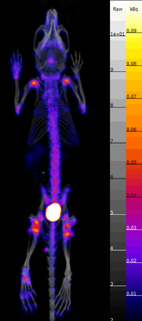
2D or 3D Scintigraphy
(SPECT: Single Photon Emission Computed Tomography)

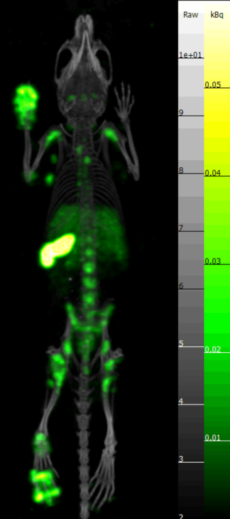
PET
(Positron Emission Tomography)

For these procedures we use gamma emitting radio-nuclides.

Principal radio-elements authorised in the imaging facility: Technetium 99m, Indium 111, Iodine 123, Gallium 67, Krypton 81m.

PET implements radio-elements called positron emitters. It is based on the detection of two 511 KeV gamma photons emitted 180° from one another when a positron is annihilated with an electron.

Radio-elements authorised in the imaging facility: Fluorine 18, Carbon 11, Copper 64.

Bio-distribution by positron emission tomography of 18FDG (fluoro-deoxyglucose) in a model of peritoneal carcinosis in the rat © TAAM
Scintigraphy of bone metabolism at 99mTc / Simultaneous bio-distribution of liposomes labelled at 111In
© TAAM

Fields of application by scintigraphy and PET

Physiological functions of the main organs

Specific applications

  • Kidneys: glomerular and tubular function
  • Lungs: perfusion, mucociliary clearance, alveolar–capillary barrier
  • Digestive tract: gastric emptying and transit
  • Liver: hepatocyte, biliary and phagocytic function
  • Hematopoietic organs (spleen, bone marrow)
  • Skeleton: osteolysis and osteogenesis balance
  • Assessment of therapeutic activity in cancer and inflammation
  • Bio-distribution and pharmacokinetics (molecules, genes, antibodies, nano-drugs)
  • Imaging of expression of αvβ3 integrins
  • Multimodal imaging coupled to in vivo bioluminescence or fluorescence and microscopy
  • Monitoring of efficacy of intrapulmonary administration (aerosols)
  • Targeting and vectorisation imaging (molecules, genes and antibodies)
  • Homing and traffic of immunocompetent and cancer cells
  • Imaging of recruitment, phagocytosis and apoptosis of macrophages
  • Imaging of angiogenesis, apoptosis and tumour hypoxia
  • Bio-distribution and pharmacokinetics

Equipment

Our equipment is as follows:

  • 2 mini gamma cameras for spatial localisation of photons (Gamma Imager, Biospace and IRIS camera, Ariès)
  • PET (eXplore Vista DR, GE). In vivo resolution: 1.2mm.
  • 1 SPECT/CT (NanoSPECT/CT, Mediso RS2D). In vivo resolution: 0.8mm.

– Monitoring, synchronising on ECG and respiration (external sensor) and maintaining body temperature (SA Instruments system)

– Containing animals under gas anaesthesia during examination, with possibility of exploring 3 mice simultaneously.