Photoacoustic and ultrasound imaging

Principle

Ultrasound scanning

Photo-acoustic imaging

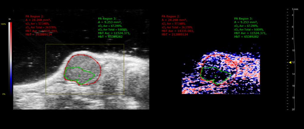
An ultrasound scan is a procedure involving the reflection of ultrasound waves, which mechanical vibrations are causing pressure variations in the host medium. An ultrasound scan makes it possible to visualise mainly soft tissues. It is also possible to obtain 3 – dimensional images when the probe is attached to a stepper motor.

When a laser is used in the form of light pulses, the energy absorbed by the biological tissues produces a micro-heating of the medium, generating the production of an acoustic pressure wave. This acoustic wave spreads and can be recorded by an array of ultrasonic transducers.

Ultrasound imaging of a cross-section of a mammary tumour (left) obtained in B-Mode. Photo-acoustic imaging of haemoglobin oxygen saturation of the same tumour (right). The zone outlined in green corresponds to a hypoxic area.
© TAAM

Fields of application

Ultrasound scanning

Photo-acoustic imaging

  • Anatomical soft tissue imaging (liver, spleen, pancreas, kidneys)
  • Exploration of cardiac function
  • Exploration of vascularisation (main blood vessels or in organs)
  •  Monitoring tumoral development (tumoral volumes and emergence of metastases)
  • Tumour induction guided by ultrasound scanning
  • Contrast ultrasonography
  • Hypoxia imaging
  • Lymphatic drainage imaging
  • Nanoparticle imaging
  • Image processing by spectral unmixing

Equipment

1 Vevo LAZR Imager (Visual Sonics Fujifilm)