Provision of services of optical imaging: 2D bioluminescence and fluorescence imagery
Principle
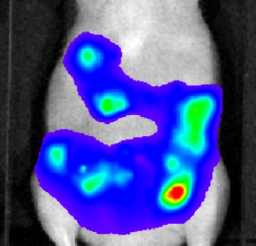
Bioluminescence
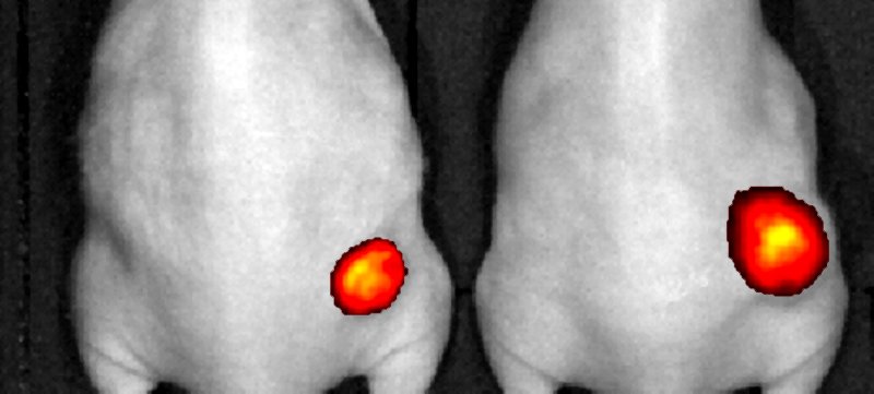
Fluorescence
This technique consists in detecting, by means of a high-sensitivity camera, the photons emitted in vivo by cells transfected or transduced with a gene encoding luciferase. This enzyme catalyses the oxidation of luciferin (injected into the animal beforehand), converting the chemical energy into light output. The imagery department possesses a bank of bioluminescent cancer cell lines but can manipulate lines developed by a client after a GMO declaration.
This imaging technique involves using molecules which, when excited by light emitted on a given wavelength, return to their fundamental state by emitting light of a slightly higher wavelength. The photons emitted inside the animal are then detected by a high-sensitivity camera. Due to the auto-fluorescence and absorption of the light by the tissues, the in vivo applications lie mainly in the near-infrared spectrum (600-800 nm).
Equipment: 2 cameras
Bioluminescence with spectral analysis and near-infrared fluorescence: IVIS Lumina and IVIS Lumina II (Perkin Elmer)